Dr. Rice has joined Beacon Orthopedics and Sports Medicine
Congratulations Dr. Rice: 2025 Cincinnati Magazine Top Doctor


Microfracture
Microfracture is a surgical technique used to repair articular cartilage damage in the knee called chondral defects. Articular cartilage is a complex avascular (no blood supply) tissue which consists of cells called chondrocytes suspended in a collagenous matrix. It appears as a smooth, shiny, white tissue at the ends of the bones which come in contact with each other to form a joint. It reduces friction when the bones glide over each other and makes the movements smooth. Alternately, it acts as a shock-absorber and enables the joint to withstand weight. Articular cartilage is subjected to normal wear and tear from daily activities and when damaged due to injury it causes pain and impaired function. Because it has no blood supply, it is unable to repair itself and can progress to arthritis. As a result, several surgical methods have been devised to restore articular cartilage and help prevent progression to arthritis.
Indications and Contraindications
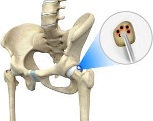
Microfracture is a common surgical technique used to repair damaged knee cartilage by drilling small holes into the knee joint to stimulate new cartilage growth.
The best candidates for Microfracture include:
- Patients whose cartilage degeneration is limited to small areas in the knee
- Younger patients who are very active and wish to return to their sport or activity
- Patients with pain in the knee from cartilage injuries
Microfracture is not recommended for patients who have widespread arthritis of the knee, are inactive, and those are unwilling or unable to participate in a rehabilitation program after the procedure.
Diagnosis
Your doctor will perform a physical examination to look for altered range of motion, swelling, and alignment of the bones. As cartilage is uncalcified it does not show up on X-rays. A high quality MRI is often required and arthroscopy is used as the final determination to what technique may be best.
Procedure
The procedure is performed under local anesthetic (numbing at the site), spinal anesthetic, or general anesthetic. Microfracture can be performed using an arthroscope, a narrow tube with a tiny camera on the end to visualize the inside of your joint. Your surgeon will make a ¼ inch incision on your knee. An arthroscope is inserted through this incision. The camera attached to the arthroscope displays the image of the joint on the monitor. A sterile solution will be pumped into your knee in order to stretch the knee and provide a clear view and room to work. Another incision is made through which specially designed instruments are inserted.
Your surgeon prepares the area by removing any damaged cartilage. Multiple tiny holes called microfractures are then made into the subchondral bone (below the cartilage) with a sharp tool called an awl. This helps to bring blood supply from the deeper more vascular bone to the surface tissues. This technique creates a nourishing environment for tissue regeneration by using the body’s natural healing abilities to form new cartilage.
Post-operative Care
Following microfracture, your doctor will recommend physical therapy to help restore motion of the operative joint. Immediately after the surgery, most patients can begin physical therapy with a continuous passive motion machine or CPM. The CPM is used to gently exercise your operated leg for 6 to 8 hours per day for several weeks. You will be instructed on using crutches to avoid weight bearing activities for a few days. You will be allowed to return to sports or other intense activities 4 months after surgery.
Risks
As with any surgery, risks are involved. Risks associated with microfracture surgery include:
- Bleeding
- Infection
- Breakdown of the newly formed cartilage may occur
- Increased stiffness of the knee joint
nodisplay
nodisplay
Hip Procedures
- Hip Arthroscopy
- Labral Repair
- Acetabuloplasty (a.k.a. "Pincer takedown")
- Microfracture
- Femoroplasty (a.k.a. "CAM takedown")
- Synovectomy
- Ligamentum Teres Tear Debridement
- Iliopsoas Recession
- Subspine Impingement Decompression
- Endoscopic Abductor Tendon Repair
- Endoscopic ITB Release
- Endoscopic Bursectomy
- Proximal Hamstring Repair
- FAI FAQs
- Ischiofemoral Endoscopic Decompression